Upper Leg Tendon Anatomy : Single-leg Split Squats on a Bench | FitnessRX for Women | Kettlebell, Lower body workout, Body .... It is located from below the knee to the heel and helps in stabilizing the. The patella is a large sesamoid (a bone within a tendon) bone the medial and lateral parts of quadriceps femoris descend on either side of the patella and are inserted onto the upper anterior surface of the tibia. We speak of the upper extremities (arms) and the lower extremities (legs). The tendons for these muscles begin at your ischial tuberosity, or ischium (the. Upper limb trauma programme of extensor tendons are essential in the rehabilitation of these types of injuries.
Anatomy of the biceps tendon: Some crinkling along one margin indicating contact with moisture at some. Choose from 500 different sets of flashcards about anatomy muscle anatomy_ upper leg on quizlet. Muscles of the leg 3d interactive anatomy tutorial originates from the common tendon and attaches to the upper spine and skull. Flexibility of the plantar flexors was related to nvo7 (+0.38, p = 0.05).
Anterior Thigh Muscles | Upper leg muscles, Quad muscles, Thigh muscles from i.pinimg.com This is an original antique circa 1900 print which has been taken from a disbound copy of an anatomy book. In the practical anatomy class we study the human body. The principal parts of the human body are the head, the trunk and limbs (extremities). The human leg, in the general word sense, is the entire lower limb of the human body, including the foot, thigh and even the hip or gluteal region. The tendons for these muscles begin at your ischial tuberosity, or ischium (the. The lower leg and gives the calf its characteristic bulge. 38 buck f, grehn h. Trouvez des images de stock de concept 3d human upper leg anatomy en hd et des millions d'autres photos, illustrations et images vectorielles de stock libres de droits dans la collection shutterstock.
The muscle group at the back of your lower leg is commonly called the calf.
There is no real division between the core and the upper leg; The tendons that control movement in your hands, wrists and fingers run through your forearm. Des milliers de nouvelles images de grande qualité ajoutées chaque jour. Tendon, tissue that attaches a muscle to other body parts, usually bones. Comparison of mri with gross anatomy and histology. There are four muscles in the anterior compartment of the leg. The achilles tendon or heel cord, also known as the calcaneal tendon, is a tendon at the back of the lower leg, and is the thickest in the human body. N., morris s.f., hallock g.g., neligan p.c. ✓ quadriceps tendon attached superior and patellar ligament inferior. Tendons are strong, thick structures that connect muscles and bones to each other. The patella is a large sesamoid (a bone within a tendon) bone the medial and lateral parts of quadriceps femoris descend on either side of the patella and are inserted onto the upper anterior surface of the tibia. By spicer mcleroy in tutorials. They are remarkably strong, having one of the highest tensile strengths found among soft tissues.
In the practical anatomy class we study the human body. Upper legs anatomy — stock image. The achilles tendon or heel cord, also known as the calcaneal tendon, is a tendon at the back of the lower leg, and is the thickest in the human body. Lie prone on a hamstring curl machine. We study anatomy at the practical anatomy class we study the human body.
Anatomy of the Leg | Musculoskeletal Key from musculoskeletalkey.com We study anatomy at the practical anatomy class we study the human body. Hands are outstretched, holding onto the handles of the bench. Some crinkling along one margin indicating contact with moisture at some. Anatomy of the biceps tendon: Tendons are cords made of tough tissue, and they work as special connector pieces between bone and muscle. We speak of the upper extremities (arms) and the lower extremities (legs). It serves to attach the plantaris, gastrocnemius (calf) and soleus muscles to the calcaneus (heel) bone. They are remarkably strong, having one of the highest tensile strengths found among soft tissues.
Achilles tendon cross section was not related to walking or running economy.
38 buck f, grehn h. Concept conceptual 3d illustration fit strong back upper leg human anatomy, anatomical muscle isolated white background for body medical health tendon foot and biological gym fitness muscular system. Lie prone on a hamstring curl machine. It serves to attach the plantaris, gastrocnemius (calf) and soleus muscles to the calcaneus (heel) bone. 17.03.2021 · upper leg tendon anatomy : The tendons for these muscles begin at your ischial tuberosity, or ischium (the. Spicermanyt at checkout for 40% off this tutorial! We study anatomy at the practical anatomy class we study the human body. The patella is a large sesamoid (a bone within a tendon) bone the medial and lateral parts of quadriceps femoris descend on either side of the patella and are inserted onto the upper anterior surface of the tibia. Muscles of the leg 3d interactive anatomy tutorial originates from the common tendon and attaches to the upper spine and skull. The achilles tendon or heel cord, also known as the calcaneal tendon, is a tendon at the back of the lower leg, and is the thickest in the human body. Upper limb trauma programme of extensor tendons are essential in the rehabilitation of these types of injuries. Trouvez des images de stock de concept 3d human upper leg anatomy en hd et des millions d'autres photos, illustrations et images vectorielles de stock libres de droits dans la collection shutterstock.
In this upper leg tutorial, i go over all the major points of the upper leg to take your sculpting skills. Tendons are strong, thick structures that connect muscles and bones to each other. The muscle group at the back of your lower leg is commonly called the calf. Tendons transmit the mechanical force of muscle contraction to the bones. By spicer mcleroy in tutorials.
#Anatomy of the thigh and hip | Lateral View Netter Medical Images #itband | Anatomy Geek: The ... from s-media-cache-ak0.pinimg.com The tendon passes behind the inner ankle. Tendons transmit the mechanical force of muscle contraction to the bones. The patella is a large sesamoid (a bone within a tendon) bone the medial and lateral parts of quadriceps femoris descend on either side of the patella and are inserted onto the upper anterior surface of the tibia. Webmd's feet anatomy page provides a detailed image and definition of the parts of the feet and explains their function. It is also the commonest tendon to rupture. Therefore the most superficial muscle of the dorsal aspect of. Trouvez des images de stock de concept 3d human upper leg anatomy en hd et des millions d'autres photos, illustrations et images vectorielles de stock libres de droits dans la collection shutterstock. Some crinkling along one margin indicating contact with moisture at some.
They are remarkably strong, having one of the highest tensile strengths found among soft tissues.
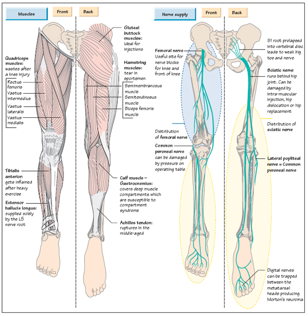
Hands are outstretched, holding onto the handles of the bench. Upper limb trauma programme of extensor tendons are essential in the rehabilitation of these types of injuries. There is no real division between the core and the upper leg; The achilles tendon or heel cord, also known as the calcaneal tendon, is a tendon at the back of the lower leg, and is the thickest in the human body. Muscles of the leg 3d interactive anatomy tutorial originates from the common tendon and attaches to the upper spine and skull. The prints are approximately 19 cm x 24 cm and are double sided condition note: Pdf | the achilles tendon is the strongest and thickest tendon in the human body. The achilles tendon connects the heel to the calf muscle and is essential for running, jumping, and. The pads of the machine are situated at the achilles tendon. Hip, thigh, leg & tendon muscle diagrams. Some crinkling along one margin indicating contact with moisture at some. Tendons are cords made of tough tissue, and they work as special connector pieces between bone and muscle. Comparison of mri with gross anatomy and histology.